







For eons, neuroscientists, including clinicians such as neurosurgeons and neurologists have striven to remedy maladies of the brain. The uniquely complex anatomy and physiology of the brain compared to other organs of the body have been a general impediment to progress. Of great interest has been failure to recover lost function after injury or damage to the brain like that which occurs with trauma or stroke. Simply put, unlike most other tissues in the body, brain cells lack the ability to regenerate; once destroyed, they are gone forever. This is referred to as a lack of “plasticity.”
Brain disorders like those resulting in abnormal movements of the extremities associated with Parkinson’s disease are ironically best treated by destroying highly specific small areas in the brain. This to some extent corrects the imbalance between related areas of the brain caused by the disease, thus ameliorating some of the symptoms. This generally requires invasive techniques with associated risks, thus there has long been interest in techniques for modulating communication between areas of the brain without opening the skull (i.e., non-invasive techniques). Focused radiation has a long and successful history in treating brain disease, but mostly for the destruction of brain tumors, and ionizing radiation has some downsides. More recently, innovative practitioners are exploring the use of focused high-intensity ultrasound energy, and even more recently, magnetic energy, to modulate connections in the brain in hopes of remedying certain brain maladies. These energies have essentially no downsides in human tissues.
Since the magnetic energy is passed into the brain through the skin and skull without incisions or bone removal, it is non-invasive and thus called “Transcranial Magnetic Stimulation.” Some of the earliest explorations of the use of this technology have involved rather diffuse magnetic fields passed through the brain not focused on any specific brain area in an effort to improve the brain’s ability to recover from traumatic injury. The idea is to alter brain cells and their connections to improve plasticity, yielding a better opportunity for repairing damage (“healing”). Recent advances in multiple technologies have further honed the potential for magnetic energy to treat certain brain dysfunction due to disease or injury.
A most recent and exciting development is that of the “connectome,” which has greatly improved understanding of brain anatomy as it relates to brain function. This is supported by two other technologies: the electron microscope and artificial intelligence. The connectome, coupled with yet two additional modern technological advances allows for the application of transcranial magnetic energy to brain dysfunction. They are “stereotactic focusing” and “Magnetic Resonance Imaging (MRI scans).” Stereotaxy uses concepts identical to those used by global positioning systems (GPS) to precisely identify and target areas of concern in the brain shown on an MRI scan. Coupling the MRI scan with the connectome allows for the identification of specific connection pathways in the brain that have been damaged by a stroke, for example, so that magnetic energy might be focused there in hopes of increasing plasticity and allowing the damaged brain to heal.
Let’s “listen in” as Chet Cooper and I chat with Dr. Zucker, a neurosurgeon working with a team in Florida under the brand BrainLink to apply these techniques in the treatment of patients recovering from stroke.
Chet Cooper: Can you give a little bit about your medical background?
Dr. Lloyd Zucker: Sure. I did undergrad at Hopkins, did my Medical School at Rutgers, neurosurgical residency at Yukon–finished in ’88. Went into private practice, left private practice after a couple of years, did a complex spine fellowship, went back into practice, then decided I wanted to pursue or expand some of the functional neurosurgery I did as a resident, but didn’t want to leave practice again, so I found a colleague that was located in Florida and basically every Friday for six months, drove and did DBS (deep brain stimulation) with him, then drove back. So started me down the path of deep brain stimulation, did that for a batch of years. There was a radiologist in our town that bought a focused ultrasound unit from Insightec, and the neurosurgeon that was working with him left, so they came to our program and said, “Would you like to learn how to do focused ultrasound?” so about five years ago, I started into focused ultrasound. Actually, our site was the lead recruiting site for two of the major Parkinson’s studies. Now we’re doing some low-frequency focused ultrasound. We’re opening the blood-brain barrier. Actually, if you’ve watched “60 Minutes” at all in the past year, one of the studies that we’re in is the same study that Ali Rezai at West Virginia presented on “60 Minutes.” That led me into working with a couple of very interesting companies in brain tractography. The final endpoint is working with a company called Omniscient. A neurosurgeon by the name of Michael Shughrue started that, and it took the human connectome project and mapped it to the brain and gave us functional connectome. So now we do resting state MRIs, and we see disorders in the brain circuits, and you can take transcranial magnetic stimulation, and you can modulate the circuits. We’ve started down the road with some interesting things. We do induce neuroplasticity for stroke patients and try and increase their rehab potential or take them if they’re plateaued, get them to a better point. I’m starting into some work on concussion patients. This is stuff that our colleagues in China, Australia, Germany are doing. Unfortunately, we’re behind in the U.S., and so we made a decision to see what was going on. A lot of our psychiatric colleagues are using TMS for depression, but not mapping the brain. We said patients need access to this technology. We began a clinic. It’s unfortunately still off label, but we’re staying out of the psychiatric lane. We’re not doing any depression or anything like that, but we think it’s not taking anything away from the psychiatrist. We think helping rehab stroke patients or concussion patients and trying to see if we can have any effect on Alzheimer’s is potentially far more important. So that’s what we do.
Dr. Tom Chappell: So, psychiatry Alzheimer’s sounds a little more of a diffuse issue, but you’re talking about mapping the brain. Are we focusing the electric magnetic radiation in any way?
Zucker: Yes. So whether we use focused ultrasound in the studies or the clinic where we’re using TMS, it’s exactly that. It is targeted. We are actually finding a site within the brain that–let’s use Alzheimer’s as an example–there are a few targets within the brain that appear to be majorly affected in the memory circuits, precuneus, a couple of other areas, and we actually stereotypically target. We get a functional MRI. We identify the target, and then we use stereotaxy to actually navigate the TMS to that target.
Products & Services


Chappell: I want to come back to the stereotaxy, but before I do, what are the psychiatrists doing? Are they targeting an area?
Zucker: I guess the best way to say that is some are not. Some are targeting directly. So, they identified the motor strip and they measure forward to the dorsolateral prefrontal cortex, or they use one of the frontal EEG sites. A small handful are starting to truly navigate using specific targets that have been identified, utilizing this new software as being an area that is dysfunctional.
Chappell: That sounds like more superficial sites, but you alluded previously to more deep sites. Are you able to reach those deeper sites with transcranial magnetic?
Zucker: Transcranial magnetic is going to be good from–depending on which one you use–between 2cm and 3 cm. Going much deeper than that, it’s either not going to be well-focused or we’re going to have to use another energy source, but with the new software that we’re using from Omniscient, we’re finding that there are a number of cortical targets that are involved in the memory circuits, so they are accessible to TMS.
Chappell: But previously, you alluded to deeper targets for Alzheimer’s. Are you reaching those with the magnet?
Zucker: There are two very recent papers that just came out of France that are reaching the deeper targets with TMS.
Chappell: How can they be sure of that?
Zucker: Well, they are using an EEG-based system, so they are actually mapping once they stimulate the EEG power and modulating it. I honestly don’t have anything other than their recently published paper, as I’m sure you can imagine, they have not let their entire secret sauce out. We’re reading a little bit into it, but our software basis and theirs is a little bit different. We’re looking at the brain slightly differently than they are. We can look at different nodes or different components of the memory circuit, and some of those are obviously not as deep.
Chappell: Are you able to objectively identify effects of the magnetic radiation? Probably not a good way to call it, the magnetic stimulation.
Zucker: Well, when you say objectively, I mean, to me, objectively in the patient–let’s say in an Alzheimer’s patient–is going to be the same thing that we’re doing in our focused ultrasound studies. An MMSC, an ADAS-COG, basically faded memory analysis because I think not using those when those are obviously the basis of what a lot of other studies had been based on wouldn’t make sense.
Products & Services
Chappell: So clinical improvement, but it’s not any way to, I don’t know, use a functional MRI or PET or SPECT or something to see changes in the brain after this treatment? There’s no way to do that.
Zucker: Since we are looking at the brain, we are basically doing a functional MRI, but I think differently than the functional MRI that you and I thought of when we were earlier in our practices. Functional MRI then was a task-based MRI. Basically, tap a finger, look at a picture, do whatever, and look at the bold signal. This is a resting state MRI, and the resting state MRI is looking at the interconnection of all of the different areas. The basic map that it’s based on is no longer broadening, it’s the human connectome atlas. There’s 180 different parcellations in each hemisphere, so 360 total. They’ve gone ahead and used a machine learning algorithm that basically is looking at all of those areas and can look and figure out. They have a normative group or a normative atlas that they basically drew from a large group that is in England. They compare to that in any area of the brain that is hypo-connected or hyper-connected, three standard deviations or more outside of the norm is termed anomalous. It can identify how many anomalous connections there are and whether they’re hypo or hyper connected. Then that’s how we start to develop the targeting because we know what the area is anatomically. Then we get an idea what it is doing physiologically and work to alter it.
Chappell: Those serve as targets, but has anyone or you guys been able to see post-treatment changes in the pre-treatment findings on those studies?
Zucker: Well, the post-treatment changes can be viewed as changes in the network maps that we have gotten pre and post. I guess you’d call it anatomic or physiologic changes as a result of the treatment, but It depends on what you’re terming the “physiologic change,” because if the resting state MRI has changed, by definition, there’s been a physiologic alteration.
Chappell: What stereotaxy technique do you use? How are you focusing this?
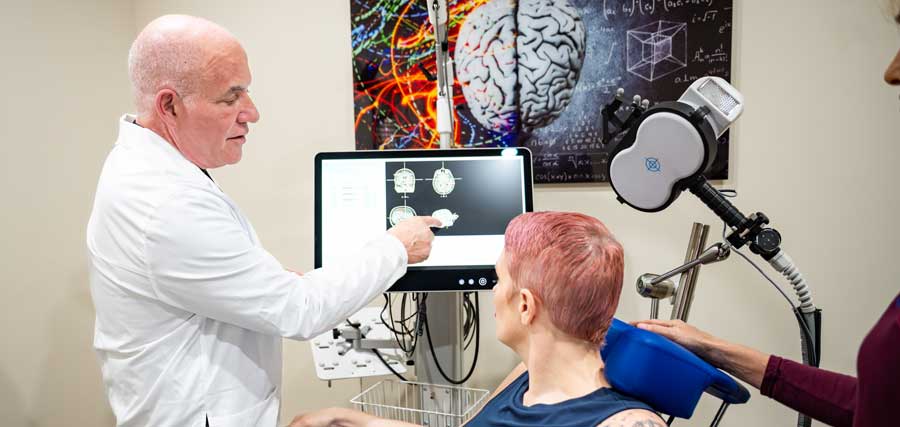
Zucker: Well, The stereotaxy outwardly is the same stereotaxy that you and I use in the OR. It’s a frameless stereotactic unit. A MRI is taken of the patient–the target that we’ve identified on the MRI is developed from the patient’s own MRI. When the patient is placed in the TMS chair, then the marker is attached to the forehead, just like we would in the OR or somewhere else, so there’s a stable point of fixation. The outside of the skull or skin is traced or identified by an infrared or laser scan. It is co-registered to the MRI. Then you have basically the stereotactic camera. So the head’s basically stereotactic space.
Chappell: Yeah, and then you have a magnetic generator of some sort that’s able to focus on that.
Zucker: Yeah, it’s a figure eight coil, so it’s not as finely focused. I do DBS, and I do focused ultrasound. I can tell you the DBS–when I do subthalamic nucleus, obviously, I’m dropping it into something that’s 4 millimeters wide and about 7 millimeters deep. The accuracy in the RSC or the spill of the TMS is not quite as good as that, but the energy field itself can be demonstrated. So, you can see where the zone of the highest intensity is and then it drops off like isodose curves on radiation.
Chappell: So you’re actually reaching those depths then with the transcranial magnetic device?
Zucker: Again, we’re not going for a brainstorm. We’re going cortical and just subcortical. To us, the power that you would have to generate to go deep, which is possible, might have deleterious or, let’s say, unpredictable effects at that cortical mantle.
Products & Services


Chappell: Just a strange thought here, and I know we want for this to be non-invasive if possible, but would it be of any benefit, do you think, just with your familiarity with magnetic stimulation, to do it through a bur hole?
Zucker: The interesting thing is the degree of absorption of the skull of the TMS is actually not that bad. Focused ultrasound, the degree of absorption by the skull is actually significant. And since ultrasound in a lot of situations can’t be directly transmitted, you need a conductive thing. So, in what we do, the head is actually in a water bath. There are other things. There are certain gels that are being used, and one device, actually, they do a craniectomy and implant the transducer in the place of the bone that’s removed. But as you said, you’re getting progressively more invasive.
Chappell: I wonder to what extent the Europeans even have been able to do this, but has anybody been able to correlate clinical improvement with any objective change in your mapping?
Zucker: Actually, some of the most in-depth work is coming out of China. Interestingly, I found out through some people that I’ve been working with in this arena, a two-tier system, even in China, of medicine. There’s this medical city that is a whole holistic, but within this is TMS as well, where they pay $80,000. Basically, it’s like this holistic Mayo clinic, but they have been doing work in strokes, work in mental health, and they are quantifying outcomes. The group in France with Alzheimer’s is quantifying outcomes. You’re going to start to see a lot more in the neuromodulation arena, I think, in general, because it’s really bursting onto the scene from a neurosurgical point of view. Prior to this, as you know, it was really in the hands of psychiatrists. They were obviously having effects in treatment-resistant depression, but thankfully, they weren’t trying to do anything else out of their lane. And it leaves a lot for us to explore.
Chappell: How do you feel about the scientific rigor in some of those settings, such as the Chinese medical city, where you have to pay to get in?
Zucker: Well, I’ve looked at two of the studies that have come out of there, and at least outwardly, placebo-controlled, randomized, blinded study. So outwardly looks well-designed. Obviously it would need to be scrutinized further, but it wasn’t a single arm, it wasn’t open label, so they’re doing it the right way to certainly try and make data that makes sense. I think you and I both know the next step is it reproducible by groups other than theirs, but they are certainly doing that. The groups in Australia have been a little bit more loosey-goosey about it. The group in Germany is doing it the right way as well. I think you’re going to see more and more well-designed studies that are making it into press. The French paper on Alzheimer’s last year, I think, was published in Brain. Obviously, if we’re getting into peer-reviewed major journals. I think the trick there, I think both of us would agree, is it needs to make it into a peer-reviewed journal.
Products & Services

Chappell: Yeah, that’s what I was getting at. Are the studies out of China peer-reviewed in a good journal like that?
Zucker: The ones out of France are peer-reviewed and have been published. Off the top of my head, I cannot tell you where the Chinese study was published.
Chappell: I know I’m beating a dead horse here, but I want to make sure I’m understanding it clearly. Is it possible or has it been shown that there’s correlation between clinical improvement and some objective change on your mapping imaging studies?
Zucker: Well, so if you’re saying to me, if I took one of my stroke patients that we’ve treated and seen a clinical improvement in the patient, measurable improvement, and seeing any change in MRI that I do to map out her circuits, pre and post-treatment? The answer to that question is yes.
Chappell: Okay. That would be, obviously, I guess somewhat helpful. One would then ask–and of course, this is where randomized controlled trials come in–just to make sure that those weren’t improvements that the patient might have experienced as a natural course of the disease.
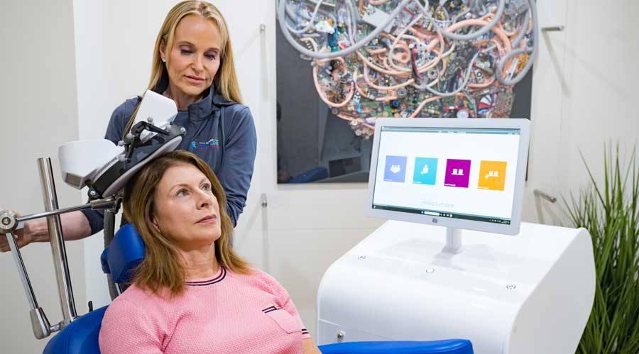
Zucker: I think that is an excellent question. The patient that I’m referencing, this patient was near a two years status post-stroke and had a moderate hemiparesis, but had more of a gait initiative variation issue where she almost described it as having to say she had to think, “I want to move my leg.” and moved her leg in that fashion. She used an assistive dog at all times for ambulation. Basically, the only demonstrable thing was a small stroke that was basically called basal ganglia. To me, it was a little bit more deep coronary modula. However, she had some very interesting changes on our omniscient side, so on the RMI, the resting state MRI, we did TMS to three separate cortical targets. We were using what’s called theta burst. So, the patient would get basically a series of TMS treatment, so five in one day. Each theta burst treatment would be followed by a period of targeted rehab and then about a half hour of rest. They would do that five times, and they did it every other day for two weeks. At the end of two weeks, she was demonstrably better. I know where your mind is going to go, hopefully. I will tell you, I have seen her at one month and at three months, and her improvements are stable and holding.
Chappell: Do you think there’s any effect from just more intensive therapy going on there, more intensive physical therapy?
Zucker: I think initially, that was definitely one of my questions. Although she was in fairly regular therapy anyway. I think ours was more intensive than what she was doing. However, in the interim between treatment and now, and the last time I saw her was about a week ago, the level of intensity of any exercise or therapy has basically been back to what she was normally doing. And so whatever gain she got from that two-week period is holding. The interesting thing is both our group and other groups that are doing this are finding out or are seeing that the changes that are induced by neuromodulation are not transient. I think where Dr. Chappell’s questions were going, which are very excellent, is, are we able to demonstrate an underlying anatomic or physiologic change in the brain where something is actually obviously going on or else the changes would not remain.
Chappell: I think you’d agree we would need rigorous controlled studies to be confident that some other effect has not taken place in the course of your treatment.
Zucker: I basically would tell you that the ideal–which would be easy to design but hard to do–is what we did for one of our focused ultrasound studies. Patients had the head frame put on, went into the magnet, and the magnet made noise, and they got no energy. So they basically got a sham treatment.
Chappell: Yeah, and then still getting the therapeutic interventions throughout the day, the physical therapy, et cetera, at the same pace and rate. To have it properly controlled, right?
Zucker: Yeah.
Products & Services

Chappell: Two other questions came to mind. Severity of impairment. So, you think you’re going to get as much of an effect, an improvement, relatively speaking, with someone with severe deficits versus less severe deficits?
Zucker: My honest opinion would be no. Our experience when talking to Dr. Sughrue, he has basically said, you can’t make a tract where there is none. If a stroke has destroyed a majority of the corticospinal tract, then no, you’re not going to get something going. However, if there’s corticospinal tract remaining and a cortical anomaly can be demonstrated, our experience so far in patients that we’ve treated is a few of the ones with the more severe deficits. Although I have been able to demonstrate strength improvement to a physical exam, I would have to say the way I feel about them is I don’t think that they have functionally improved to a point where the strength improvement has translated into a function benefit for them.
Chappell: Rang bells of my master’s thesis many, many years ago–the debate over the mechanisms of recovery of function after brain damage, whether there’s plasticity for new pathways in a higher mammal, or rather, our theory was that there were existing pathways that were dormant and were awakened after an injury. This would be theoretical, of course. Do you have an idea as to what you think is going on at the cellular level?
Zucker: I believe that there’s got to be something going on at the cellular level because I have a hard time understanding how some of these improvements remain stable if there hasn’t been. I think you would very much enjoy a book that probably now came out about three months ago. It’s called Connectomic Medicine. The author is actually Dr. Sughrue. I think a lot of the very astute questions and curiosities that you’re asking and have in terms of how they got to this point and what they think the TMS is doing and how they look at the brain, I think it would fit very interestingly with your original thesis. I think it rests somewhere between the two options that you offered. I don’t think pathways are dormant. I think that there are multiple nodes. I think in a very severe deficit, you’ve basically damaged a major node. If there’s been minor nodes that have been damaged, you can potentially push messaging over a pathway that not so much was dormant but wasn’t as operational in the functional task that you were of the circuit. And by activating or utilizing that circuit, that may help define what plasticity really is.
Chappell: Yeah, that’s exactly along the lines of our thesis. This was many, many years ago. A lot of stuff has been done since then. It begs the question as to whether or not there’s an actual distinction between what we’re saying there. What is plasticity after all. Back then, we thought of it as the pathways were being regenerated, and as you know, we didn’t feel like the central nervous system of a mammal could do that. But anyway, maybe we are still talking about a type of plasticity.
Zucker: Well, and the other thing, I mean, part of this software has been a surgical, stereotypic software because what the contention is, is there’s really no such thing as non-eloquent brain. And by being able to map out circuits and understand areas that I think, well, I don’t know when you finished your training, but many of us were raised in the world of pseudo-architectural brain. And this area was safe and this area is not, and don’t work that way.
Chappell: Yeah. No surprise to me, but I came along about the same time you did.
Zucker: I’ve said to people, and they find it hard to believe when I say, I’m actually in many ways relearning things about the brain, some which I took for granted and others which I’ve scratched my head. I think you and I have probably both done operations where we’ve looked when the patient’s woken up in recovery and said, “How the hell?” To be able to unlock it a little bit in advance, especially with the intra-axial brain tumors, makes it a very interesting thing to do.
Products & Services

Chappell: Yeah, let’s not go down that pathway today. No, not at all. I guess if we’re stimulating with magnets only to a depth of a couple of 3 centimeters, theoretically, we feel like that’s still modifying pathways that extend deeper into the brain, I suppose.
Zucker: Absolutely. I think truly, if you grab Connectomic Medicine–it’s on Amazon–what it will also give you is a slew of reference experiences, but it really begs that very question that you’re asking, do some of these subcortical injuries or subcortical pathways have cortical manifestations or attachments? Obviously, the cell body is where? The cortical mantle. There’s some part of the loops got to be there, but it begets, I think, the second question that you would ask me, which I’ll ask myself is–does modifying at the cortical level modify the entire circuit?
Chappell: Right. I guess you’d have to almost be assured of that if you’ve got patients who have clinically improved with deeper lesions and have improved clinically with your treatment.
Zucker: Yeah. How should I say this? I’m an optimistic skeptic. I want to continue doing more because I’d like to try and figure out why they get better. Working with some people at Scripps, which we happen to be lucky enough to have in Palm Beach County that are doing some very interesting things on a biochemical level. I’ve gone to them with what I’m doing and saying, “What do you think?” They’re great bench researchers, and they’re like, “We’d love to try and figure this out.” I might have some more answers for you in the days to come.
Chappell: Now, that touched on my other question, and that is, is there any role or even possibility for an animal model that would be of some use?
Zucker: After discussions with them, they do have some interesting animal models that I was not aware of, both in lots of arenas, the addiction arena, Parkinson’s, you name it. It’s one of those things where you get enough people from different arenas in the same room and you start them talking, you can get some very interesting things going. It’s a really interesting combination of bench academic research and clinical translation. They’re very excited about our willingness to talk to them. Conversely, I’m like, “Wait a second, guys. I’m thrilled that you’re willing to talk to us.”
Chappell: Look, it’s critical, but just occurred to me earlier, what frameless system are you using?
Zucker: The frameless system that we’re using with the TMS, it’s a company called ANT, but it looks just like what I think you and I would have used in the OR. So it has the camera with three receptors, the adhesive localizer that we put onto the forehead, has three reflectorized balls, just like the Medtronic system. The probe that we use to trace out the calvarium has the three reflectorized balls, so it’s literally a slightly smaller but intrinsically the same system that we use on our Medtronic Series 8 in the OR.
Chappell: Also not important, but you may find of some interest, I was a resident under Bart Guthrie, who, we believe, was the first person to develop a Deep Brain Stimulator. There’s some debate about that, but I worked on at least one of the first frameless systems as it was being developed.
Products & Services

Cooper: Let me step in here for a second as a layperson. You work with TMS, but also focused ultrasound. How would you choose what you’re doing? Can you say what the difference is for the readers?
Zucker: Two very different modalities. My work in focused ultrasound has been basically two different things. High-frequency focused ultrasound is ablated, so you’re actually destroying a small piece of brain. First indication that it was cleared for was unilateral essential tremor. It’s now cleared for bilateral essential tremor. Also, we’ve done work with that for Parkinson’s disease, and there’s a paper being submitted for publication on an area of the brain called the pallidothalamic tract, which is for Parkinson’s disease. Low-frequency focused ultrasound is different. It opens the blood-brain barrier. It has applications in neuro-oncology. It is being looked at at the present point in time for some uses in addiction. There are other areas of brain modulation that are being looked at because the low frequency is not destructive. It just modulates. There’s a lot of work going into that. TMS–completely different application. We’re looking at it for stroke and concussion, not for movement disorders. And given the work that just came out of France, since we’re doing low-frequency focused ultrasound for Alzheimer’s, there’s a study going on at the present point in time. We may look at some of the applications of TMS in Alzheimer’s. So, I have a healthy opinion of what a disability really is…you’re allowed to ask why.
Cooper: Well, I actually was trying to avoid that, but go ahead.
Zucker: My dad had polio at age three. He actually ended up with 21 surgeries and a trophic left leg. However, he was ambulatory without braces until he got into his early ’70s and had more orthopedic manifestations of his altered gait that became problematic. But he played handball, he played softball. He wasn’t mobile like everybody else, but he never let it stand in his way. I learned that for some people, disability is physical, for some people, disability is mental. Or a combination. Not mental as in a disease, but all you had to do is watch the Paralympics, and there were people that had outward physical disabilities that were doing shit that I couldn’t even imagine doing.
Cooper: Back to both of the ultrasound and the TMS, they’re considered non-invasive, correct?
Zucker: Correct.
Cooper: I had heard you say you were doing something with deep brain stimulation. Isn’t that considered always invasive?
Zucker: Yeah, you drill a hole, then you pass something through the brain. So, that falls into the invasive category. In fact, there were people that I saw for essential tremor that when they learned I would be drilling a hole and passing something physically through their brain, they were like “Absolutely not.” They have come back and gotten focused ultrasound. And even though I’m passing beams of energy through their brain with the express purpose to destroy a tiny area, the fact that I’m not using a scalpel, not cutting the skin, not drilling a hole, and not leaving something permanently implanted, it is far better received by those patients than the concept of how they define invasiveness.
Cooper: Sure. You talked about MRI and functional MRI. Where does that come from? Is that in your lab? Do you have a facility that actually has that equipment, or is that a hospital nearby that you’re working with?
Zucker: Well, I’m at both a freestanding MRI institution and the hospital. These are image sequences that can be done on standard 1.5 and 3 Tesla MRIs. We use a 3 Tesla MRI. Basically, it uses something called fusion tensor imaging, not trying to get too scientific, but it looks at the flow of water through the axons in the brain. There are different ways to interpret that data that allow different levels of what’s called tractography or seeing those actual axons within the brain. That’s structural. Then you can take it to a functional level, which is what this newest software is doing.
Cooper: So it’s measuring water. I always thought it was measuring heat.
Zucker: Actually, no, but when we do focused ultrasound that’s ablative, it is generating heat, and the MRI turns into the world’s most expensive thermometer.
Chappell: MRI looks at the polar shifts in water molecules?
Zucker: Exactly.
Cooper: I guess maybe because the image is red and all that, they’re saying that you can pick up the heat, the slight change.
Chappell: All that colorized stuff you see, that’s done with a computer. They tell the computer to colorize certain differences in signal and so forth.
Zucker: That’s all artificial. As neurosurgeons, when the MRI era began, we used to joke because everything that was black on a CT, they made white on an MRI and everything. They did this so it would confuse everybody but the radiologists.
Chappell: They played another little trick on us, too. They stood at the foot of the bed and put the right on the left, and we had to stand at the head of the bed and put the right on the right.
Zucker: Yeah. Sorry for all these collegial jokes that are coming out.
Cooper: Now, why the term Brainlink?
Zucker: Well, we were looking for a way to try and explain what we were doing in terms of trying to link brain function and what we were doing together and tried to come up with a host of different names. The one that was the most attractive to all of us was Brainlink.
Cooper: Did you develop the software or did another company develop the software?
Zucker: Actually, a neurosurgeon, Dr. Sughrue. If you go back about 12 or 13 years while he was practicing, and he’s still a practicing neurosurgeon, he was working at making what was called the Human Connectome Project, which was really a magnificent endeavor. But once it was finished and we had this connectome map of the brain, nobody was doing anything with it. He’s the one that took part of that along with other labs and started linking it to brain function and have developed a much more intricate map of the brain than we ever had before.
Products & Services
Cooper: It was briefly mentioned, but I don’t think the question was asked directly. Any thoughts or any work going into creating some clinical trials?
Zucker: Given the fact that a lot of the other things that I do were involved in clinical trials, the answer is yes, we’re collecting data prospectively regardless. Clinical trials are usually either investigator initiated or sponsor initiated. The unfortunate part of true IRB (Institutional Review Board) trials is they’re expensive, they’re hard to design. When you’re talking about longitudinal studies, looking at disease that needs to be measured out over a period of time, let’s say three or five years, you can see the amount of person power as well as economics tends to be difficult. They’re going to get there. It’s just taking time.
Cooper: I’m somewhat aware of the challenges of clinical trials. Anything else you would like to add?
Zucker: Well, I appreciated the opportunity to talk. I mean, obviously, this is new and different. We think it has a remarkable potential, and we’re only seeing the tip of the iceberg in terms of its applications. We’re hopeful that if we can help push the needle and move the science forward, then we’ve made a contribution.
Cooper: At this point, you’re unique in what you’re doing in Florida, or are there other facilities similar to what you’re doing with the software and the new app application?
Zucker: There is no one else in Florida, to my knowledge, utilizing this software in combination with TMS. There are one or two other sites in Florida that have the software and are using it for surgical navigation, which is the same way we started. That’s a hospital-based modality. We’re not the only place that has the software, but we are the only place that is applying the software in this fashion.
Cooper: How’s the business? Are the people aware of what you’re doing?
Zucker: It’s a good question. We intentionally started slowly. We’re being very cautious on screening patients and who we take in. The word is certainly getting out, and we’re getting more and more inquiries. We’re really very–as would be appropriate–result-oriented. We’re trying to be very particular in terms of who we choose and how we apply it. You’d love to have the greatest impact on the most severe limitation outwardly, but that’s the best to start I know everyone’s different.
Cooper: Knowing that each patient is unique and different, is there a way that you could describe what might be a typical treatment session, number of sessions, amount of time for the treatment, et cetera?
Zucker: I think that the best thing to say that is the treatments are disease-specific. One of my partners is treating tinnitus, and the tinnitus treatments in terms of the length of the treatment and how many treatments different than the stroke treatments. A classical stroke treatment patient is treated for two weeks every other day five sessions each day that they’re being treated. The length of the actual physical treatment can range between two and eight minutes. But then with the rehab and the rest period, each one of those is about an hour. Depending on how many targets we treat in the individual patient, they can be in our clinic for 5 to 8 hours on that day.

Chappell: What’s he targeting for tinnitus?
Zucker: Well, there’s a couple of different ways of looking at it. He’s looking at Heschl’s gyrus, but there’s some other non-directly auditory targets that Dr. Sughrue has developed as well.
Chappell: Any signs of hope there? That’s a dilatating condition that I see fairly frequently in my practice now.
Zucker: The hesitation you hear in my voice is that I don’t think the direct auditory targeting is going to be the solution, but I don’t think there’s been enough done in terms of treating the other areas. I can give you some anecdotal successes, but not enough to really say that I think they have the answer. The other thing that I think you may find interesting is one of the targets that’s non-auditory is an anxiety target.
Chappell: Very interesting.
Cooper: So, they’ve mapped some of this in the brain that you can target that specific area?
Zucker: Yeah.
Cooper: Interesting. And what about the psychiatrists? Are they looking and trying this as well? Do they have any outcomes?
Zucker: Most of the psychiatrists are not. They’ve been doing this for a number of years, and they’ve been doing it in a way that is not as technology-intensive. I think that’s one of the reasons that we didn’t march down that road–even though Dr. Sughrue has identified five different targets in terms of between OCD, ADD, major depressive disorder as different sites within the frontal lobe and on a temporal area. The real question that then has to be answered is, does that specificity of targeting translate into better outcomes than what the psychiatrists are already getting? We really didn’t want to get into, if you don’t mind the vernacular, a pissing contest on whose way of doing it was better because we really didn’t want to treat psychiatric disease. There’s a whole host of things, especially if you’re doing something in a psych patient and they worsen in some way, you really need to be intimately involved with the psychiatric end of things. We felt that there were enough areas like stroke, like tinnitus, like concussion that are woefully underserved that we might be able to have some impact on. So, we didn’t go down the psychiatric lane.
Cooper: In concussion, what is it that you map where the concussion–where the trauma occurred?
Zucker: We’re talking about people’s convicute and chronic concussion symptoms, that patient where the concussion is not going away. You can show with this software, again, areas of dysfunction. By trying to treat those areas, you can ameliorate some of the “post-concussive symptoms.”
Products & Services
Cooper: When you say about the mapping, you’re talking about what Dr. Chappell had said earlier about the difference in polar activities?
Zucker: Yeah, basically what an MRI is doing is it’s forcing all your protons to align in one direction. Mother nature doesn’t like that. They like entropy–disorganization. Then the MRI, basically, you are exposed to a radio wave and all of your protons flip, and as they flip back into a disorganized state–it’s simplifying it to a point–but basically, the patient becomes a radio transmitter, and there are receivers that receive that signal and computers that interpret that signal. That’s how you get the beautiful pictures that you get because things that are in a crystalline lattice–like bone, it ignores the bone–it sees what’s inside. That’s how it can peer through, and you get all these remarkable pictures that look like an anatomic atlas.
Cooper: I’ve always challenged that thought of entropy and chaos. I think it’s the opposite. I understand what you’re saying about the organization when it’s pulled some energies, causing some specific alignment for a period of time for using that energy. But I think it goes back to equilibrium, not to chaos.
Zucker: Well, and I don’t disagree you, but the equilibrium can be a balance of organization and disorganization. I don’t think it’s totally disorganized or totally organized. I don’t disagree with you. I think your theory explains my brain a lot better than one of the other polar opposites.